Structure
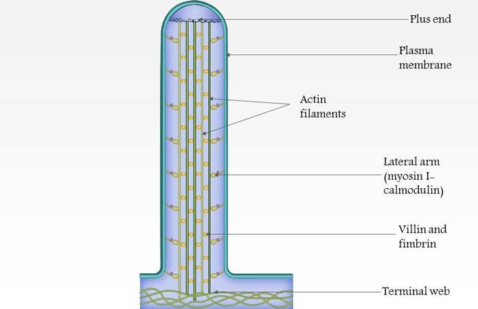
Microvilli are extensions of the cell membrane, and as such, they are linked to the structure of a cell membrane. The difference is that, while a cell has many organelles within it, a microvillus often boasts of few or none at all. The inner core of a microvillus is formed by a dense bundle of actin filaments. These 20 to 30 actin filaments are cross-linked within the microvillus by bundling proteins, including villin, espin, and fimbrin. In intestinal cells, the microvillus is attached along its length to the cell membrane extension by some lateral arms made of myosin 1a and calmodulin (a calcium-binding protein). On one side, the myosin 1a is bonded to the actin side by binding sites, while on the other side, it is bonded to the membrane by lipid-binding domains. [2]
In the formation of microvilli, the actin filaments in the cytosol are found closer to the cell membrane. These actin filaments are important for cell movement, and maintenance of shape. The cell is induced to produce microvilli that would serve a specific purpose in the cell. Nucleation occurs, and the cell surface protrudes with the actin filaments in it. The microvilli formed are usually similar in length and composition in most cell types. [3]