X-Rays
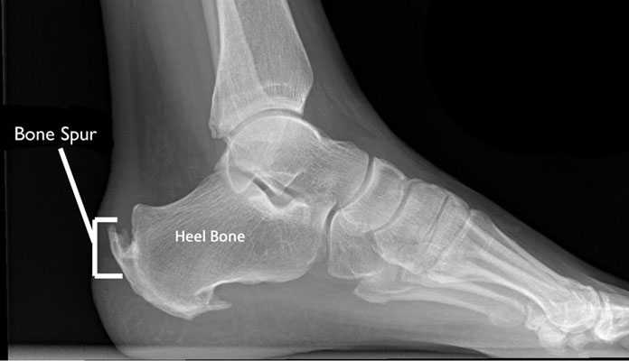
X-rays are another common imaging technique used for diagnosing Achilles tendinitis. Unlike ultrasound, it cannot produce images of tendons and soft tissues. However, this is an effective way to rule out other conditions with similar symptoms. During this procedure, you need to sit still while the doctor makes some images of your heel and leg. The results will show whether bone spurs grow in these areas or whether the tendon is calcified or thickened. X-rays can also identify the exact location of the inflammation, thus allowing for a better treatment plan.[3]