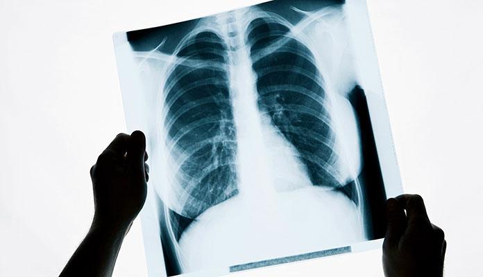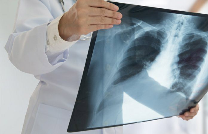
Chest X-Ray
A chest X-ray is another way to diagnose emphysema. It is basically a radiographic exam of the diaphragms, ribs, large arteries, heart, and lungs. It can be done at your bedside, in a doctor’s office or a radiology lab. In most cases, your doctor might complete an initial chest X-ray to give you an overall diagnosis and then do it regularly during the treatment to monitor the overall progress. During an X-ray, your emphysematous lungs might look hyper and normal markings of your blood vessels might be less prominent. In addition, the diaphragms can be flattened because of the hyperinflation in your lungs. A major drawback of this method is that changes in X-rays cannot be noticed until your emphysema is rather extensive. In addition to helping with diagnosis, a chest X-ray can also rule out other similar conditions like lung cancer. [4]