
Chest X-ray & CT of the lungs
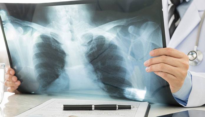
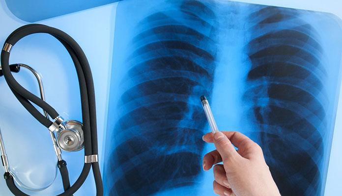
Chest X-ray is one of the diagnostic means for pneumonia, and it allows radiologists to see your lungs directly. During the procedure, you will stand in front of an X-ray machine with your upper body undressed. Your radiologist might ask you to change position for a different view of your lungs. There are black-and-white images (chest radiographs) produced after your chest X-ray exam. The findings of pulmonary infiltrate on your chest radiograph might represent possibilities of pneumonia or tuberculosis. Another way to diagnose pneumonia is CT scans. Many people are confused between X-ray and CT, and they do have similarities in mechanisms and imaging. X-ray is a conventional way to examine bones, cancers, and pneumonia. CT is a more sophisticated technology with powerful X-rays. It detects not only dense substances but also soft tissue, muscles, or other body organs. Hence, doctors can see your lung more clearly on a CT scan.